Video-Film
Dr. Jean Hissette's Research Expeditions to Elucidate River Blindness
This film has been completely remade in 2010 using Hissette's own celluloid material from ~ 1935 as well as material from the film "Harvard African Expedition of 1934", Archives of the Harvard University Medical School, followed by a short presentation of a focal TV program from Belgium in 2005 (TV Lux).
This film is presented on DVD in the annexe to the book (same name), Kaden 2011
The first of 5 parts of the film can be viewed here.
The basic material was:
Harvard African Expedition of 1934 (original)
Six reels of 35 mm safety film B & W (total film running time = 54.2 minutes) from this Harvard African Expedition have been found in archives of the Harvard University Medical School, rediscovered in Boston in 2002 with the assistance of the science correspondent Dr. Dr. Roland D. Gerste, Gaithersburg, Washington D.C. These films are transferred to DV CAM (Digital Video); scenes were identified with the aid of the notes made by Richard Pearson Strong.
Hissette's own original film material
Since about 1935, the material had been thought to be lost but not long ago it turned up as a VHS-video (running time = 30 minutes) and finally only the original celluloid film itself remains missing. This old celluloid film material had been recorded with two different early hand-cranked movie cameras. One was a heavy Zeiss camera with an axial optic for image control and the other one was a lightweight AGFA Box with an image control via an optic through a magnifier viewed from above with the camera held at waist level.
Belgian TV Lux-television contribution The Belgian TV Lux-television contribution on Florenville in 2005: During the "Cercle archéologique et historiques de la region de Florenville" in 2005 Edouard Hizette from Florenville, Belgium, organized an exhibition as well as presentations on Dr. Jean Hissette, the son and general practitioner of the city.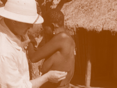
Fig. 34 (Chapter IV): Hissette is demonstrating filarial nodules upon the trunk of a patient,
Belgian Congo 1934 A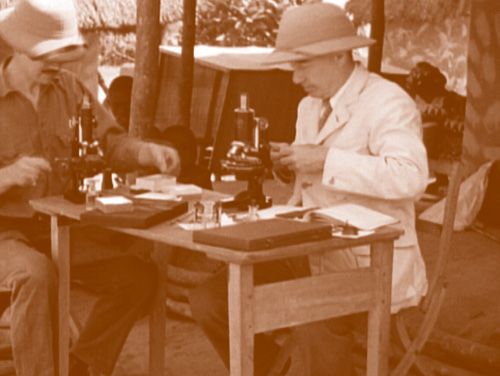
Fig. 35: Strong (right side) and Bequaert (left) at the microscopes, Belgian Congo 1934